atle.dev
pausedI-JEPAViTself-supervisedmedical imaging

I-JEPA on histology images

Image JEPA (ViT, block masking) pretrained on PathMNIST at 224px on a 24 GB Mac, then frozen-feature probes against a random-init encoder to measure what self-supervision actually buys.

WhenSep 2026
Headline78.6% · best linear probe (random-init encoder: 74.3%)
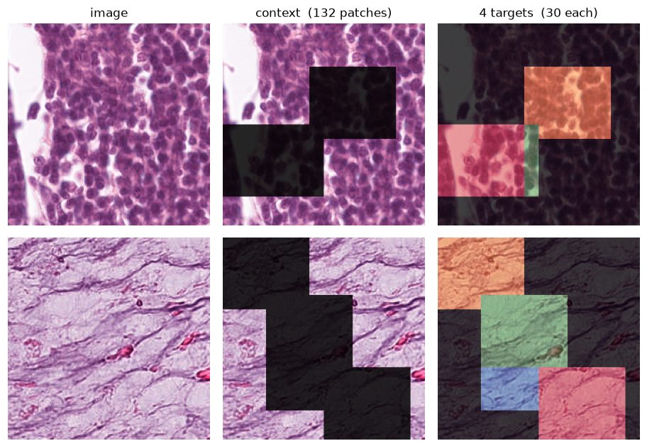
PathMNIST tiles with I-JEPA context and target block masks
I-JEPA masking on PathMNIST tiles. Left: image. Middle: the context the encoder sees (132 patches). Right: four target blocks the predictor must predict in latent space.

Why

Ball-in-cup taught the action-conditioned side of JEPA. The other big use case is a self-supervised visual backbone for domains with many images and few labels, like pathology. I wanted to see it work at a realistic resolution on my own hardware: PathMNIST at 224 px, ViT encoder, block masking, no EMA target.

Engineering on a 24 GB Mac

The 224 px split ships as a compressed .npz, and loading it pulls ~13.5 GB into RAM, which put training straight into swap. Decompressing each split once to an uncompressed .npy and memory-mapping it brought data RAM to roughly zero. It also let multiple data-loader workers share pages instead of copying.

Probing the frozen encoder

Frozen features, 9 tissue classes, 7,180 test images. The same probes run on a randomly initialised encoder as a baseline:

Random-init encoderlinear probe
74.3%
I-JEPA, epoch 1linear probe
77.2%
I-JEPA, epoch 4linear probe
78.6%
Random-init encoder5-NN
63.1%
I-JEPA, epoch 45-NN
78.1%

The linear-probe gain over random init is real but modest. The nearest-neighbour gain (+15 points) is the clearer sign that the representation organises tissue types. The features' effective rank also roughly doubled between epochs 1 and 4.
Lesson A random-init baseline is essential. A linear probe on random ViT features already reaches 74%, so an absolute number on its own says little about what pretraining did. Longer runs weren't automatically better either: probe accuracy has to be tracked across checkpoints, not just read at the end.
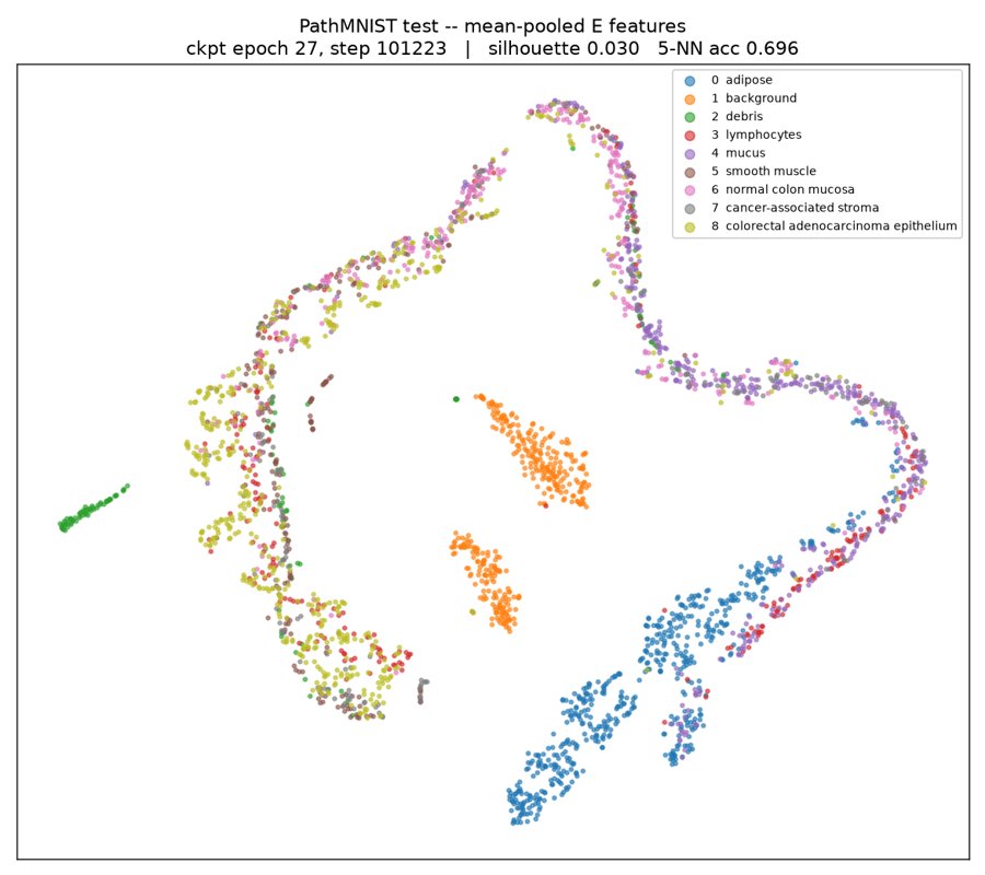
2-D embedding of mean-pooled encoder features coloured by tissue class
Mean-pooled features from a later checkpoint, projected to 2-D. Background, adipose and debris separate cleanly. The epithelial and stromal classes stay entangled.